
It took a educated eye to see the abnormality on the affected person’s X-ray. There, hidden behind the acetabulum was the shadow of a small bone spur.
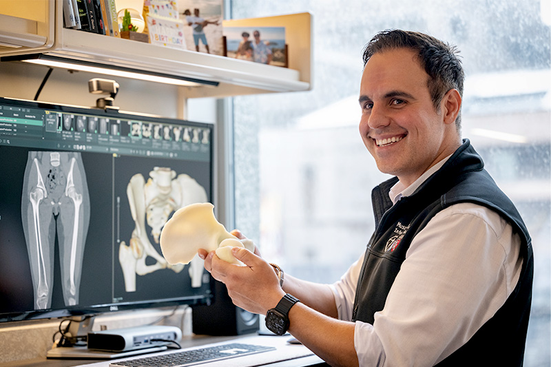
“At first look, this seems like a standard, wholesome hip,” says Young-Jo Kim, MD, PhD, director of the Child and Young Adult Hip Preservation Program at Boston Kids’s Hospital. “However right here you possibly can see the tip of the bone spur behind the joint — which is uncommon.” Kim goes on to level out that the socket is overlaying the femoral head greater than typical, however that too is delicate, one thing a much less skilled orthopedist might simply miss.
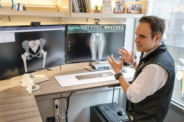
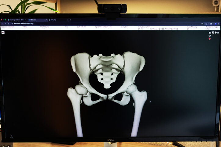
When he pulls up a 3D mannequin of the identical hip, the problems turn into far simpler to identify. The bone spur that was nearly imperceptible on X-ray is clearly seen, as are crimson areas indicating areas of bone impingement. Kim factors to a inexperienced line on the femoral head. “That’s usually the place the sting of the socket needs to be,” he says. As a substitute, the road is nicely contained in the socket.
For the previous a number of many years, 3D photographs have helped orthopedic surgeons visualize advanced orthopedic circumstances. They’ll additionally assist youthful surgeons extra precisely establish musculoskeletal points and sufferers perceive the supply of their ache. Time and price, nonetheless, have been limiting elements. Usually, an imaging technician generates 3D photographs from MRI or CT scans. Relying on the division’s workload, this will take per week or longer.
3DModelerTM, a brand new innovation at Boston Kids’s, might change this image.
Well timed, cost-effective entry to 3D imaging
3DModeler is an extension of VirtualHip, a software program program developed at Boston Kids’s that uses 3D imaging and artificial intelligence (AI) to assist the analysis and therapy of hip circumstances. After its profitable rollout in 2023, leaders within the Orthopedics and Sports Medicine Department needed to make 3D imaging a regular of care in each space of orthopedics and sports activities medication. To take action, they’d want a whole-body 3D system.
The aim was formidable. Ata Kiapour, PhD, director of Boston Kids’s Musculoskeletal Digital Innovation & Informatics Program describes the challenges. “As a tertiary referral middle, Boston Kids’s treats lots of of sufferers with uncommon and complicated circumstances,” he says. Not solely that, however given the hospital’s pediatric inhabitants, some sufferers’ bones are smooth and immature, whereas others are absolutely ossified.
The system would subsequently should be sturdy sufficient to mannequin advanced structural abnormalities whereas taking the maturity of a given affected person’s bone under consideration. To be most helpful, it will additionally want to provide these fashions shortly and at minimal value.
The group took benefit of latest advances in AI fashions and infrastructure to fulfill every of those challenges. “We educated an AI mannequin to create 3D geometries of bones at totally different phases of growth,” says Kiapour. Additionally they educated this system to precisely mannequin sufferers’ smooth tissues.
A transparent view of cartilage
Kiapour pulls up a 3D mannequin of a knee with osteochondritis dissecans. Utilizing the 3DModeler, he rotates the picture, declaring bones and tissues.
“Right here you possibly can see the distribution of cartilage,” he says, indicating the bottom of the femur. “If there’s harm, you possibly can see that.” And certain sufficient, close to the joint, a small lesion is seen the place a bit of cartilage has separated from the bone. Whereas an skilled radiologist or surgeon would seemingly have seen the lesion on MRI, it’s way more seen, significantly to an untrained eye, on the 3D mannequin.
“We had been capable of create this mannequin from an MRI inside 5 minutes utilizing 3DModeler,” says Kiapour. As soon as the system is up and working, clinicians will have the ability to generate 3D fashions from their computer systems and handheld gadgets — no imaging technician wanted. In the event that they need to print a 3D mannequin, for instructing or patient-education functions, the computer-generated fashions can assist this.
A 3D future for orthopedics
As of now, 3DModeler can generate 3D fashions of any a part of the physique from a CT scan. If the unique picture is an MRI, the software program can at the moment create 3D fashions of a affected person’s hips, knees, or ankles. Work is underway to broaden this record to incorporate the entire physique as nicely.
3DModeler was launched for beta-testing in Boston Kids’s Orthopedics and Sports activities Medication Division this summer season. A interval of gathering clinician suggestions is underway, and the group expects that inside a yr, 3D modeling will likely be a regular a part of medical apply for Boston Kids’s orthopedic sufferers, supporting medical resolution making for physicians in any respect ranges of experience.
“Having a greater visualization software improves each analysis and surgical planning,” says Kim. “You possibly can see very clearly what the issue is and how you can repair it.”
And whereas AI made the expertise of 3DModeler doable, having medical leaders keen to spend money on non-traditional analysis was simply as crucial.
“Due to the imaginative and prescient of our senior leaders, together with Dr. Kim, Boston Kids’s often is the solely orthopedic division on the earth to have deployed one thing like this in our clinics,” says Kiapour.
Be taught extra in regards to the Musculoskeletal Digital Innovation & Informatics Program, Child and Young Adult Hip Preservation Program, and Orthopedics and Sports Medicine Department.
Trending Merchandise


The Pout-Pout Fish

Giraffes Can’t Dance


Moo, Baa, La La La!

Manhattan Toy Skwish Color Burst Rattle ...

Doggies